Services

Pigtail for liver abscess, Pleural effussion, ascites & pancreatic Pseudocyst

A pigtail catheter is a medical device that is used to drain fluid from various parts of the body. It is called a pigtail catheter because it has a curled end, which resembles the tail of a pig.

Pigtail catheters can be used to drain fluid from the liver in cases of liver abscesses.

Pigtail Catheter Placement for Liver Abscess, Pleural Effusion, Ascites, and Pancreatic Pseudocyst:

A pigtail catheter is a flexible, small-caliber tube with a curled end that can be inserted into various body cavities or organs to drain fluids or collections, such as abscesses, pleural effusions, ascites, and pancreatic pseudocysts. Here’s an overview of pigtail catheter placement for these medical conditions:


1. Liver Abscess Drainage:

  • A liver abscess is a collection of pus within the liver. Drainage is necessary to remove the infected material and promote healing.
  • Drainage helps relieve symptoms, such as pain and fever, and aids in the administration of antibiotics.

2. Pleural Effusion Drainage:

  • A pleural effusion is an accumulation of excess fluid in the pleural space around the lungs. It can cause breathing difficulties and discomfort.
  • Drainage of pleural effusion improves breathing and relieves chest discomfort.

3. Ascites Drainage:

  • Ascites is the abnormal accumulation of fluid in the abdominal cavity, often associated with liver disease or other conditions.
  • Ascites drainage helps alleviate abdominal distension and discomfort.

4. Pancreatic Pseudocyst Drainage:

  • A pancreatic pseudocyst is a collection of fluid and tissue debris around the pancreas, often caused by pancreatitis.
  • Drainage of pancreatic pseudocysts reduces symptoms, such as abdominal pain, and allows the pseudocyst to heal.

FNAC and Biopsy

Fine-Needle Aspiration Cytology (FNAC) is a diagnostic procedure used to investigate lumps or masses. In this technique, a thin, hollow needle is inserted into the mass for sampling of cells that, after being stained, are examined under a microscope. The sampling and biopsy considered together are called fine-needle aspiration biopsy (FNAB) or fine-needle aspiration cytology (FNAC).

FNAC and biopsy are both diagnostic procedures used to determine the presence of cancerous cells in lumps or masses. FNAC involves the use of a thin needle to extract cells from the mass, while biopsy involves the extraction of sample cells or tissues for examination. Both procedures are commonly used in the diagnosis of cancer and other diseases.

1. FNAC (Fine Needle Aspiration Cytology):

  • Indications: FNAC is primarily used for evaluating superficial lumps or masses, including those in the breast, thyroid, lymph nodes, and soft tissues. It is often employed to diagnose the nature of the tissue, such as whether it is benign or malignant.
  • For –
      1. Breast Lump/Masses
      2. Enlarged Lymph Node
      3. Oral Cancers/Masses
      4. Thyroid Nodules/ Lump

2. Biopsy

  • Indications: Biopsies are used to diagnose a wide range of conditions, including cancer, infections, and autoimmune diseases. They can be performed on various body parts, including skin, organs, and mucous membranes.
  • For –

1. Lung Cancer
2. Breast Cancer
3. Liver Cancer
4. Abdominal Cancer/Lump
5. Prostate Biopsy Cancer (TRUS)
6. Bone Lesions

Colour Doppler & Ultrasound Facilities

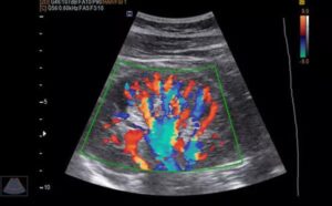
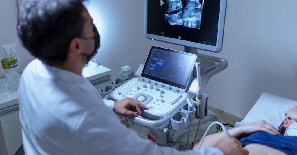
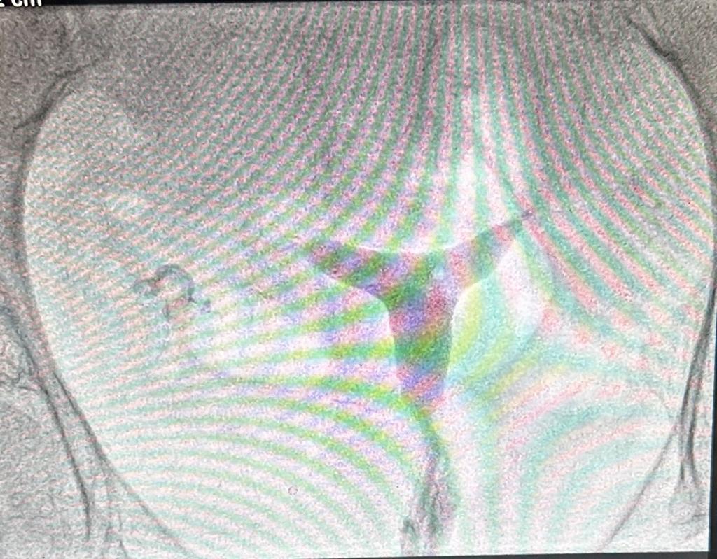
Color Doppler is a technique used in medical ultrasound that allows for the visualization of blood flow direction and velocity within a user-defined area. This is achieved by color-encoding Doppler information and displaying the colors as an overlay on the 2D image of the heart. Color flow Doppler is used frequently in sonography to semiquantitatively assess overall blood flow to a region of interest. Depiction of the general velocity and direction of blood flow within the heart and blood vessels is of primary importance in echocardiography and vascular ultrasound respectively.

Ultrasound facilities that offer Color Doppler services are equipped with specialized ultrasound machines that are capable of performing this technique. These facilities may be found in hospitals, clinics, or standalone imaging centers. One such facility is Atulaya Healthcare, which offers Color Doppler Ultrasound services at their centers in Chandigarh, Jammu, Faridabad, and Ludhiana. They use the Acuson X500 ultrasound system from Siemens, which offers versatility for today’s multispecialty applications. Doppler ultrasound images can help the physician to see and evaluate blockages in blood flow (such as clots), narrowing of vessels, tumors, and congenital malformation.

Introduction: Color Doppler and ultrasound facilities play a crucial role in modern healthcare. These diagnostic tools utilize ultrasound waves to create real-time images and assess blood flow. Below, we explore the facilities, applications, and benefits of Color Doppler and ultrasound in healthcare.

1. Definition:

  • Color Doppler Ultrasound: A medical imaging technique that combines traditional ultrasound with Doppler technology to visualize blood flow within the body’s blood vessels. It uses color-coded images to display the direction and velocity of blood flow, aiding in the diagnosis of various medical conditions.

2. Facilities:

  • Ultrasound Machines: These devices use high-frequency sound waves to create images of internal structures. They are equipped with Color Doppler capabilities to assess blood flow.
  • Dedicated Ultrasound Labs: Healthcare facilities typically have specialized ultrasound rooms with state-of-the-art equipment.
  • Portable Ultrasound Units: Smaller, portable ultrasound devices are available for use in different clinical settings, including emergency departments, operating rooms, and remote locations.
  • Skilled Sonographers: These healthcare professionals are trained to operate ultrasound machines and perform exams.

3. Applications:

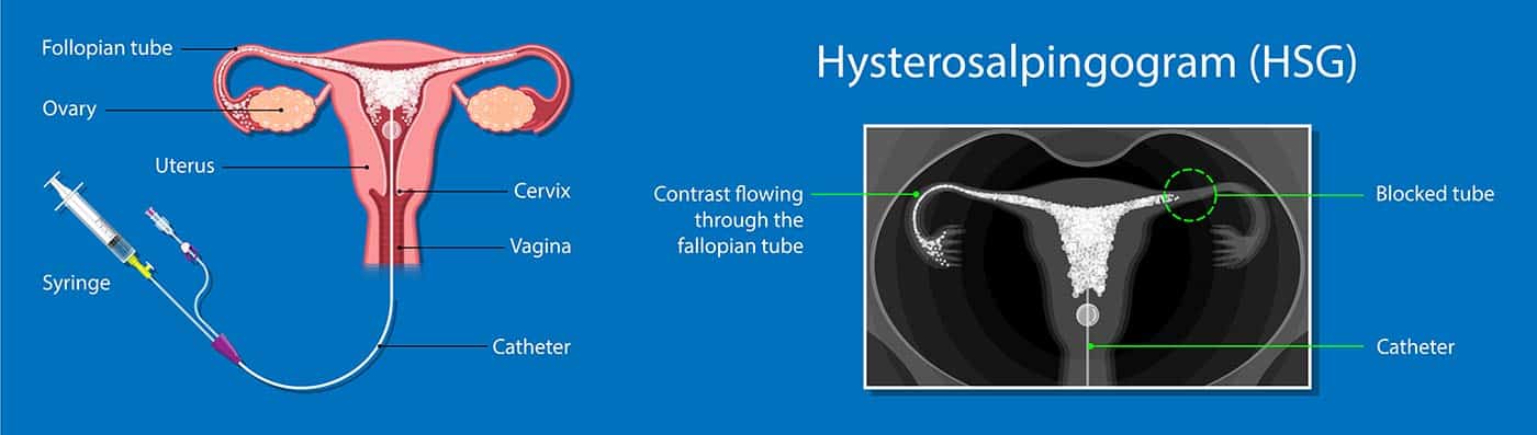
  • Obstetrics and Gynecology: Used for prenatal care, monitoring fetal development, and detecting gynecological conditions.
  • Cardiology: Assesses heart function, blood flow, and identifies heart conditions.
  • Abdominal Imaging: Evaluates the liver, kidneys, pancreas, and other abdominal organs.
  • Vascular Studies: Examines blood vessels for blockages, clots, or abnormal blood flow.
  • Musculoskeletal Imaging: Assesses muscles, tendons, and joints for injuries or diseases.
  • Emergency Medicine: Rapid assessment of trauma patients, including detecting internal bleeding or organ damage.
  • Urology: Evaluates the urinary tract and assists in diagnosing conditions like kidney stones.

4. Benefits:

  • Non-Invasive: Ultrasound and Color Doppler are non-invasive techniques, meaning they do not require surgical procedures or radiation exposure.
  • Real-Time Imaging: Provides immediate, dynamic images, allowing for quick diagnosis and treatment decisions.
  • Safety: Ultrasound is considered safe for all age groups, including pregnant women and infants.
  • Versatility: Used in various medical specialties, making it a versatile diagnostic tool.
  • Cost-Effective: Generally more affordable than other imaging modalities like MRI or CT scans.

5. Limitations:

  • Operator-Dependent: The quality of images and accuracy of diagnosis can depend on the skill and experience of the operator.
  • Limited Penetration: Ultrasound has limited penetration capabilities, making it less effective for imaging deep structures or obese patients.
  • Not for All Conditions: While highly useful, ultrasound may not be the best choice for certain conditions that require detailed imaging or functional assessment.

Conclusion: Color Doppler and ultrasound facilities are essential components of modern healthcare. They provide valuable insights into a wide range of medical conditions while offering safety, versatility, and cost-effectiveness. However, their effectiveness can be operator-dependent, and they may not be suitable for all clinical scenarios. When used in conjunction with other diagnostic tools, they contribute significantly to accurate diagnoses and patient care.

BRTO, PARTO, TIPPS

Balloon-occluded retrograde transvenous obliteration (BRTO) is a technique used by interventional radiologists in the treatment of gastric varices, particularly those with prominent infra-diaphragmatic portosystemic venous shunts (e.g. gastro-renal and gastro-caval shunts).

  • The technique is more popular in Asia, where it is a first-line treatment for gastric variceal hemorrhage.
  • The modified versions include balloon-occluded antegrade transvenous obliteration (BRTO), coil-assisted retrograde transvenous obliteration (CARTO), and vascular plug-assisted retrograde transvenous obliteration (PARTO).
  • The advantages of PARTO include decreased procedure time, decreased post-procedure monitoring and removes the risks associated with an indwelling balloon catheter, i.e. rupture.
  • TIPS (Transjugular Intrahepatic Portosystemic Shunt) is another valuable option in controlling esophageal (when endoscopy has failed) and gastric variceal bleeding. TIPS has proven very effective in acutely controlling both esophageal and gastric variceal hemorrhage.

Signs & Symptoms: BRTO is primarily utilized to address the following symptoms and conditions:

  • Gastric Varices: Swollen veins in the stomach that can lead to potentially life-threatening bleeding.
  • Portal Hypertension: Increased pressure in the portal vein, often associated with liver cirrhosis, which contributes to the development of gastric varices.
  • Bleeding: Gastric varices can cause symptoms such as vomiting blood or passing bloody stools.


Investigation and Diagnosis

  • Endoscopy
  • Imaging Studies

Available Treatments:

  1. Balloon-Occluded Retrograde Transvenous Obliteration (BRTO)
  2. Endoscopic Therapy
  3. Transjugular Intrahepatic Portosystemic Shunt (TIPS)


PICC line Placement

A Peripherally Inserted Central Catheter (PICC) line is a long, thin tube that is inserted through a vein in the arm and passed through to the larger veins near the heart.

A PICC line is commonly recommended for cancer treatments, liquid nutrition (total parenteral nutrition), infection treatments, and other medications that can irritate small veins. Once your PICC line is in place, it can also be used for

  • Blood draws
  • Blood transfusions
  • Receiving contrast material before an imaging test.

PICC Line Placement: A Comprehensive Guide

A Peripherally Inserted Central Catheter (PICC) line is a long, thin, flexible tube that is inserted through a peripheral vein, typically in the arm, and threaded into a larger vein in the body, usually near the heart. PICC lines are commonly used for various medical purposes, including the administration of medications, chemotherapy, intravenous (IV) fluids, and for blood draws. Here’s a detailed overview of PICC line placement:


1. Indications:

  • Long-Term IV Therapy
  • Chemotherapy
  • Antibiotics
  • Nutrition
  • Frequent Blood Draws

2. Placement Procedure:

  • Preparation: Before the procedure, the patient’s medical history is reviewed, and any contraindications are assessed. The insertion site is typically in the upper arm.
  • Ultrasound Guidance: Often, ultrasound is used to locate the appropriate vein for insertion.
  • Sterile Technique: The procedure is performed under sterile conditions to reduce the risk of infection.
  • Local Anesthesia: A local anesthetic is used to numb the area where the PICC line will be inserted.
  • Insertion: A healthcare provider inserts the PICC line through a small incision, typically in the arm, and guides it into a larger vein, usually the superior vena cava, using imaging guidance.
  • Catheter Securement: The PICC line is secured in place with sutures and adhesive dressings.
  • Chest X-ray: A post-placement chest X-ray is often done to confirm the correct positioning of the PICC line.

3. Care and Maintenance:

  • Dressings: The site where the PICC line enters the skin is covered with a sterile dressing, which should be kept clean and dry.
  • Flushing: The line is flushed regularly with sterile saline or heparin to prevent blockages and maintain patency.
  • Dressing Changes: Dressings are changed regularly to prevent infection.
  • Monitoring: Patients with PICC lines are monitored for signs of infection, blockage, or other complications.
  • Removal: When the treatment course is completed or if complications arise, the PICC line is removed by a healthcare provider.

4. Patient Education:

  • Patients with PICC lines are educated on proper care, signs of infection, and what to do in case of complications.

5. Removal:

  • PICC lines are removed when they are no longer needed, or if complications arise, by a healthcare provider.

Overall, PICC line placement is a valuable medical procedure that provides long-term access for various treatments and therapies while minimizing the need for repeated needle sticks. However, careful care, monitoring, and attention to potential complications are essential for the safe and effective use of PICC lines.

Embolization of Intracranial Aneurysm

Embolization is a minimally invasive procedure used to treat aneurysms by filling them with material that closes off the sac and reduces the risk of bleeding. It is performed from “within” the artery (endovascular) through a steerable catheter inserted into the bloodstream and guided to the brain.

Embolization of brain aneurysms and arteriovenous malformations (AVM) uses imaging guidance to place small, soft metal coils into an aneurysm to block the flow of blood and prevent the aneurysm from rupturing. It also is used to fill AVMs – abnormal connections between arteries and veins – with liquid embolic agents (similar to fast-sealing glue).


Embolization of Intracranial Aneurysm

Embolization of intracranial aneurysms is a minimally invasive medical procedure aimed at treating abnormal bulges or weak areas in blood vessels within the brain. This technique involves the insertion of tiny coils or devices into the aneurysm to block blood flow, thereby preventing the risk of rupture. Below, we provide a detailed overview of this procedure, including its definition, indications, procedure, and potential benefits and risks.

2. Indications:

  • Intracranial Aneurysms
  • High-Risk Aneurysms

3. Procedure:

  • Angiography
  • Endovascular Embolization
  • Follow-up Imaging

4. Benefits:

  • Minimally Invasive
  • Rupture Prevention:
  • Preservation of Brain Function

HSG with opening of Tubal Blockage (FTR)

Hysterosalpingogram (HSG) is an X-ray procedure that is used to view the inside of the uterus and fallopian tubes. It is often used to see if the fallopian tubes are partly or fully blocked.

  • Fallopian tube recanalization (FTR) is a nonsurgical procedure to clear blockages in the fallopian tubes.
  • The procedure is done under sterile conditions to avoid the possibility of introducing infection.
  • The overall time to do the procedure is about a half an hour, most of which is spent in getting you comfortable, readying the equipment, and cleaning the skin.
  • The actual time spent in evaluating and unblocking the tubes is usually only a matter of minutes and you will be able to leave about a half hour after your procedure is completed.
  • The success rate of reopening blocked tube(s) is 95%.


1. Hysterosalpingography (HSG):

HSG is a radiological test that involves the injection of a contrast medium (dye) into the uterine cavity through the cervix. X-ray images are then captured to visualize the uterine and tubal structures, allowing for the assessment of their patency and any abnormalities.

done for-

  • Infertility
  • Tubal Blockage

2. Fallopian Tube Recanalization (FTR):

FTR is a procedure aimed at reopening blocked fallopian tubes to restore fertility. It can be performed in conjunction with HSG.

  • FTR is typically recommended when HSG identifies tubal blockages as a potential cause of infertility.

MWA ( Microwave ablation for liver Tumor, uterine fibroids )

Microwave Ablation (MWA) is a minimally invasive medical procedure used for the treatment of various medical conditions, including liver tumors and uterine fibroids. It employs microwave energy to heat and destroy abnormal tissues. Below, we provide an in-depth overview of MWA, including its definition, indications, procedure, and potential benefits and considerations for both liver tumors and uterine fibroids.

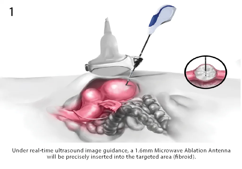
1. Microwave Ablation (MWA):

Definition: MWA is a medical technique that employs microwave energy to generate heat within tissues, causing thermal ablation or destruction of targeted cells.

Indications:

    • liver
      • hepatocellular carcinoma (HCC)
      • metastases from colorectal cancer
    • pancreas
      • advanced and non-resectable pancreatic tumors – pain control
    • kidney
      • small renal masses
    • lung
      • lung tumors
    • bone
      • painful metastatic lesions
      • osteoid osteoma

Procedure: During MWA, a thin, microwave-emitting probe is guided into the target tissue using imaging guidance, such as ultrasound or CT scans. Once positioned, the microwave energy is delivered to heat and ablate the tissue, causing coagulation and cell death.

Advantages
    • over radiofrequency ablation (RFA)
      • less heat sink effect
        • allowing efficient ablation close to large vessels
      • propagate into fat and calcifications
        • leading to homogeneous coagulation necrosis
      • consistently higher intratumoral temperatures
        • cause larger and fast necrosis
      • faster ablation times
        • lower doses of sedatives might be required
      • larger tumor ablation volumes

In conclusion, Microwave Ablation (MWA) is a minimally invasive procedure used to treat various medical conditions, including liver tumors and uterine fibroids. Its effectiveness and suitability depend on factors like the type, size, and location of the condition being treated. Patients should discuss their specific case and treatment options with their healthcare providers to determine the most appropriate approach.

TACE ( Transarterial Chemoembolisation ) for Liver Tumor

Transarterial chemoembolization (TACE) is a minimally invasive treatment for liver cancer. It involves injecting a combination of cancer-fighting drugs and an embolic agent into the tumor to block its blood supply, causing the cancer cells to die. This procedure is also known as chemoembolization.

TACE is a specific type of chemoembolization that blocks the hepatic artery to treat liver cancer. Liver cancer tumors can grow new blood vessels (called angiogenesis), which get most of their blood supply from the hepatic artery, while the rest of the liver tissue gets blood from the portal vein. Because of this, doctors can block the hepatic artery to cut off the blood supply to the tumor without affecting the rest of the liver as long as blood is normally flowing toward the liver in the portal vein.


Transarterial Chemoembolization (TACE) for Liver Tumors

  • Transarterial Chemoembolization (TACE) is a medical procedure used to treat liver tumors, particularly hepatocellular carcinoma (HCC) and, in some cases, metastatic liver tumors.
  • TACE combines chemotherapy and embolization techniques to target and reduce the blood supply to the tumor while delivering a high concentration of anticancer drugs directly to the tumor site.
  • TACE is a minimally invasive procedure performed by interventional radiologists. It involves the selective infusion of chemotherapy drugs directly into the blood vessels supplying the liver tumor, followed by the injection of embolic agents to block these vessels,  minimally invasive

2. Indications:

  • TACE is primarily used to treat liver tumors when:
    • The tumor cannot be surgically removed.
    • The tumor is confined to the liver or is dominant within the liver.

3. Procedure:

  • Patient Evaluation: Before TACE, patients undergo imaging studies, such as CT or MRI, to assess the size, location, and vascular supply of the tumor.
  • Catheter Insertion: A catheter is inserted through a small incision, typically in the groin, and threaded up into the hepatic artery, which supplies blood to the liver.
  • Chemotherapy Infusion: Once the catheter is in place, anticancer drugs (chemotherapy agents) are infused directly into the hepatic artery. The goal is to deliver a high concentration of drugs to the tumor while minimizing systemic side effects.
  • Embolization: After chemotherapy infusion, embolic agents, such as tiny particles or beads, are injected through the catheter to block or reduce blood flow to the tumor. This helps trap the chemotherapy drugs within the tumor and deprive it of oxygen and nutrients.
  • Repeat Procedures: TACE may be performed in multiple sessions, typically at intervals of a few weeks, depending on the response and the extent of the disease.

4. Benefits:

  • TACE has several advantages, including:
    • Targeted Treatment
    • Reduces Blood Supply
    • Improves Survival
    • Minimally invasive
    • No cuts, Sutures
    • No Anesthesia Required
    • One Day Treatment