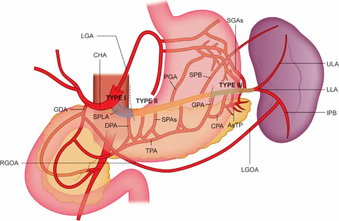
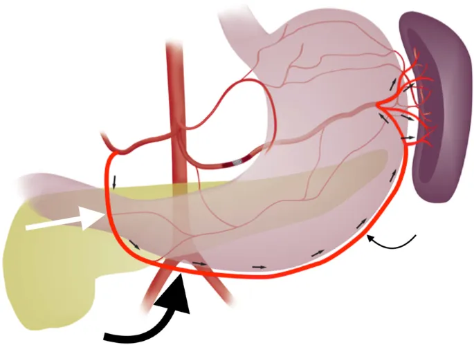
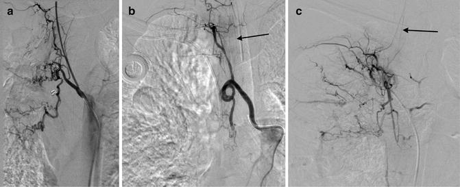
Splenic Artery Embolization (SAE)
Splenic Arterial Embolization (SAE) is a minimally invasive medical procedure used to address various conditions related to the spleen. Dr. Konika Chaudhary, an expert in this field, provides insights into this valuable procedure.
Procedure Overview
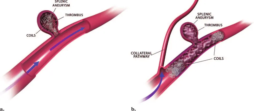
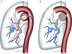
- Imaging and Assessment: SAE begins with thorough imaging to evaluate the spleen’s condition and identify issues such as ruptures, aneurysms, or vascular malformations.
- Catheter Placement: A slender catheter, guided by Dr. Chaudhary, is inserted through a small incision, reaching the splenic artery.
- Embolization: Tiny particles or coils are introduced into the splenic artery, effectively blocking blood flow to the problematic area while preserving the spleen’s overall function.
Indications for SAE
SAE is employed for various medical conditions, including:
- Splenic Trauma: It can be crucial in managing spleen injuries and reducing the risk of bleeding complications.
- Splenic Aneurysms: SAE helps prevent aneurysm rupture, a potentially life-threatening event.
- Splenic Vascular Malformations: It can alleviate symptoms and reduce complications associated with vascular malformations.
Benefits of SAE
- Minimally Invasive: SAE requires only small incisions, leading to quicker recovery times compared to traditional surgical options.
- Preservation of Spleen Function: While addressing specific issues, SAE aims to maintain overall spleen function whenever possible.
Considerations
While generally safe, SAE does carry some potential risks, including infection, allergic reactions to embolic agents, unintended vessel blockages, or damage to surrounding tissues. Discuss these with Dr. Chaudhary before the procedure.
Conclusion
Splenic Arterial Embolization (SAE), in collaboration with the expertise of Dr. Konika Chaudhary, provides effective solutions for a range of spleen-related medical conditions. If you or a loved one requires SAE, consult with Dr. Chaudhary to explore its potential benefits for your specific health situation.
Splenic Artery Embolization (SAE) Read More »

 Considerations
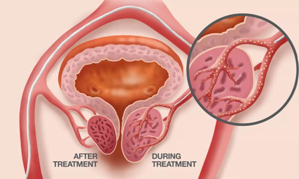


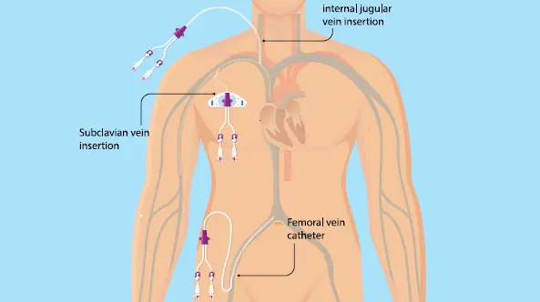
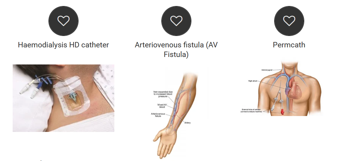
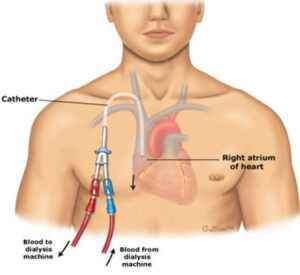 For patients suffering from kidney failure, undergoing dialysis is a lifeline that ensures the removal of waste and excess fluids from their bodies. To facilitate this crucial medical procedure, a specialized catheter known as Permacath, or Permanent Dialysis Catheter, is often recommended. Permacath is a unique solution for patients whose veins are exhausted or unsuitable for AV fistula creation, as it offers several advantages over other dialysis access methods. In this article, we will delve into the benefits of Permacath and provide insights from Dr. Konika,Renowned interventional radiologist, about its placement and care.